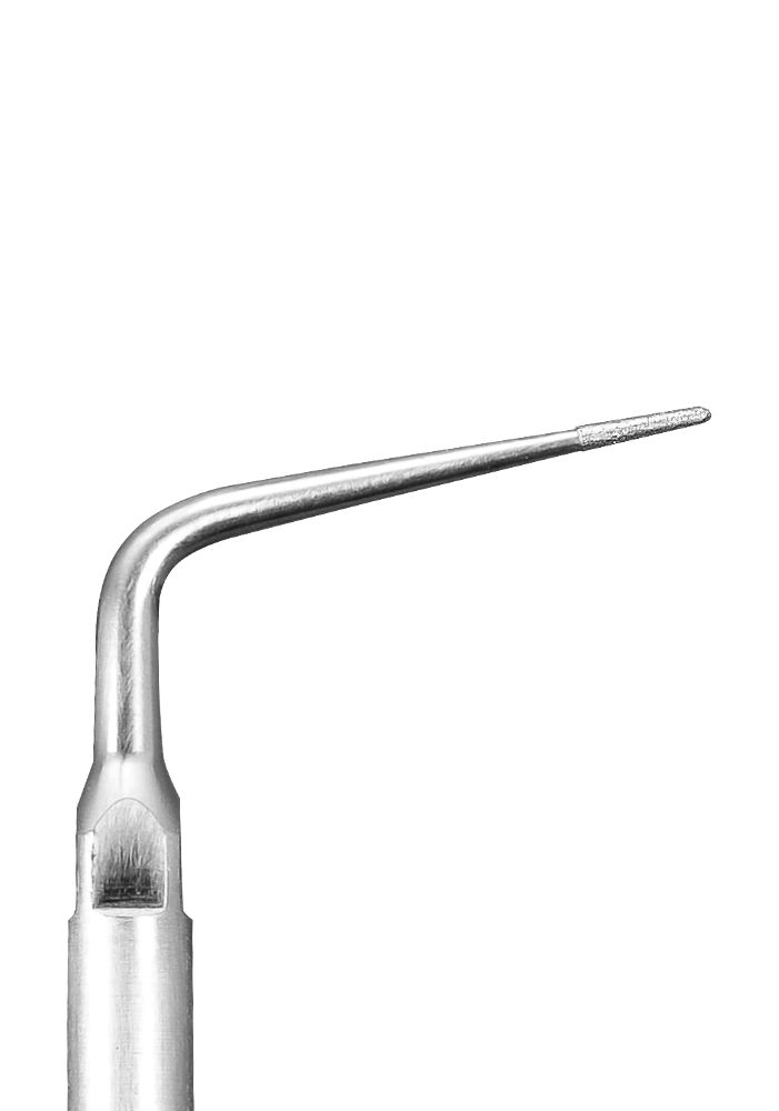
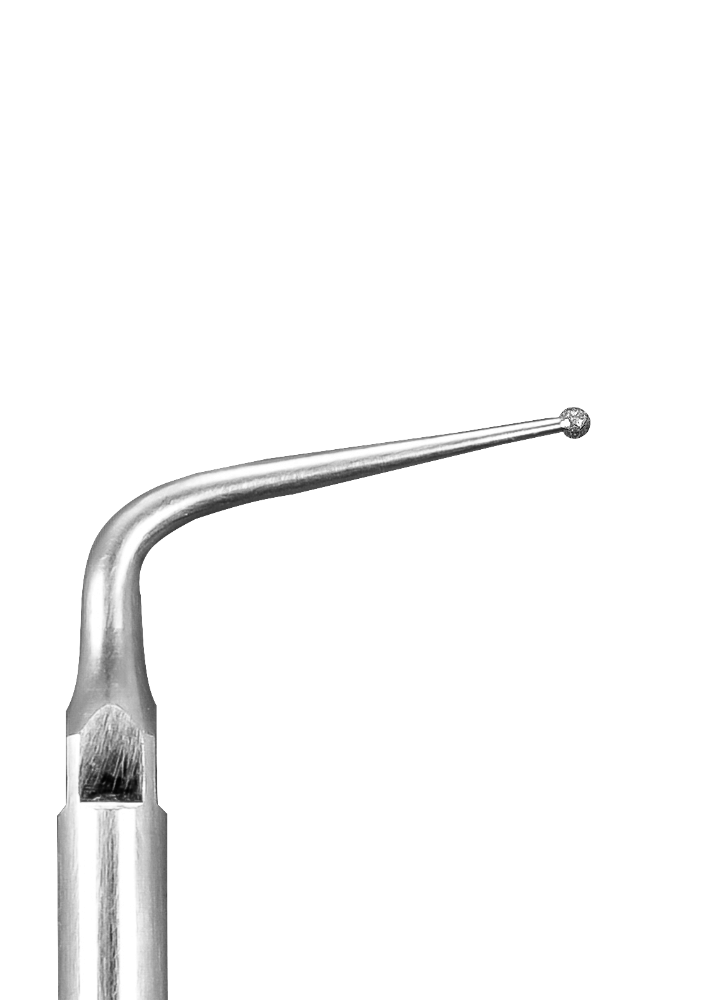
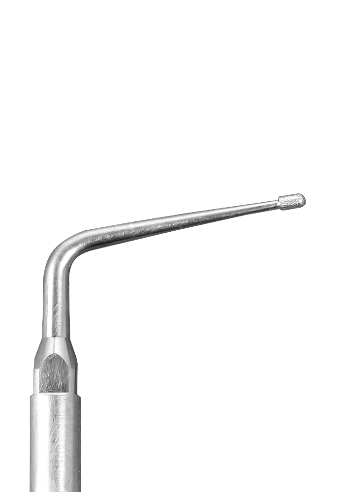
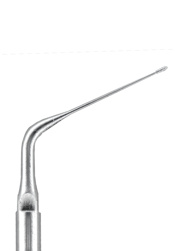
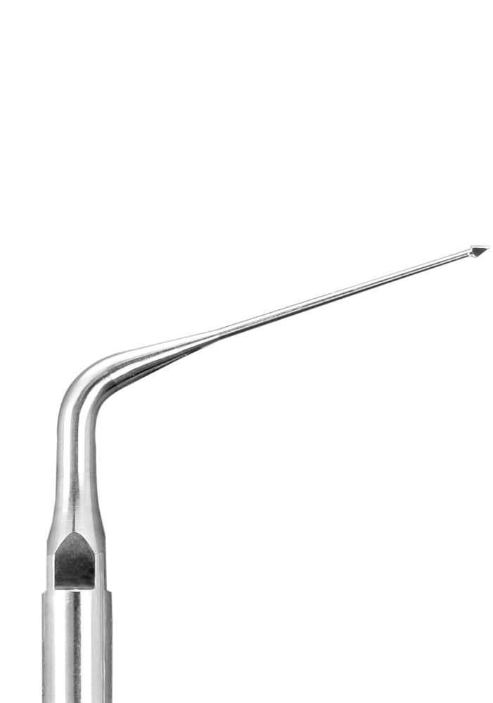
Locating Second Mesiobuccal Canals (MB2)
Leave No Canal Untreated with Ultrasonics
Studies on maxillary first molar anatomy show that the second mesiobuccal canal (MB2) is present in 65-90% of the cases. Ultrasonic instrumentation with a diamond coated insert (E2D or E6D) along the line connecting the primary mesiobuccal and the palatal canals will often reveal the MB2 orifice location. Clinically, the use of magnification and ultrasonics is the most effective technique to locate the MB2 canal.
Step-by-Step Calcified Canal Location
1 – Using a diamond coated ultrasonic insert (E2D or E6D), create a groove connecting the MB1 and the palatal canals. This will remove secondary dentin, lighter in color.
2 – The dentin covering the pulp chamber floor and its mesial wall must completely removed.
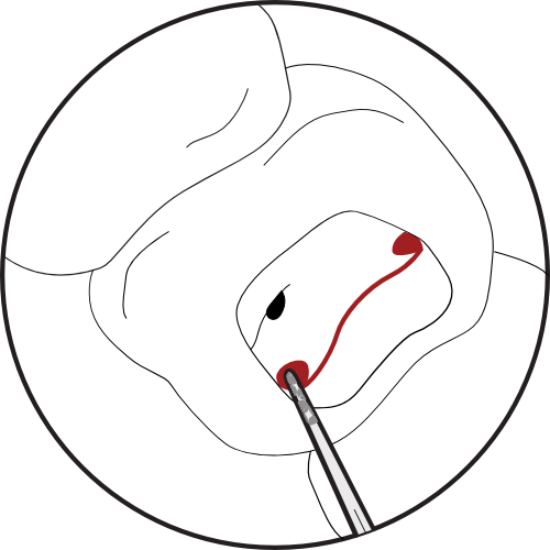
1. Create a groove to connect the MB1 and the palatal canal.
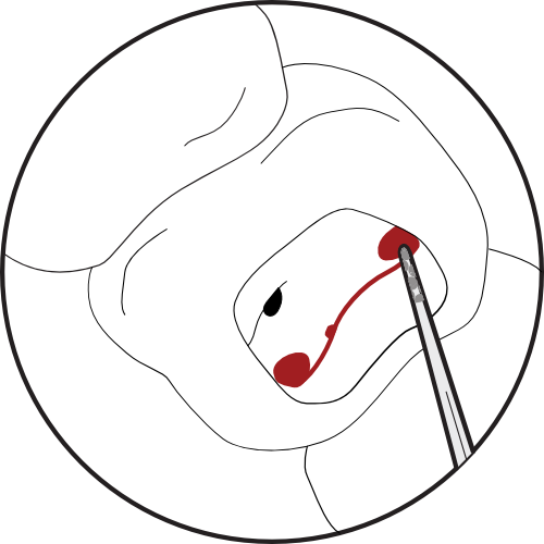
2. Locate the MB2 orifice along the groove.
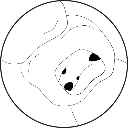
3. Use an appropriate file to negotiate and prepare the canal.
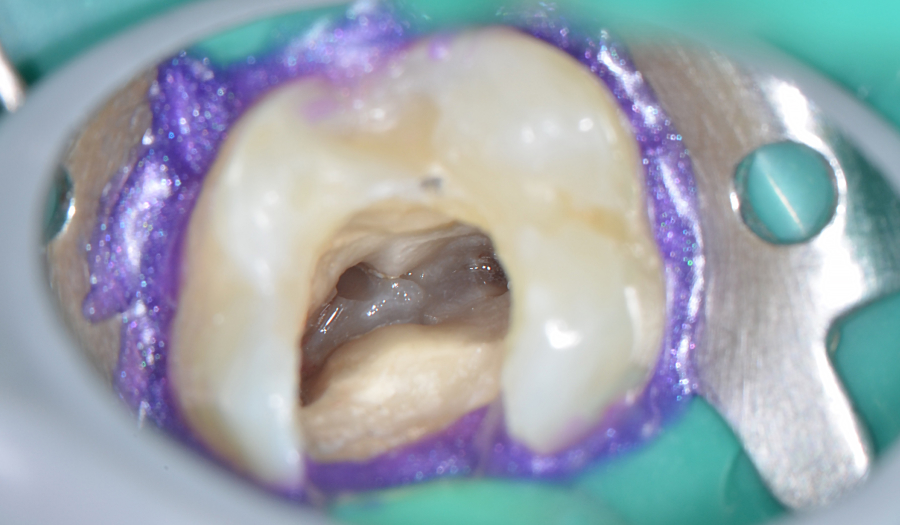
Pulp chamber of a maxillary first molar.
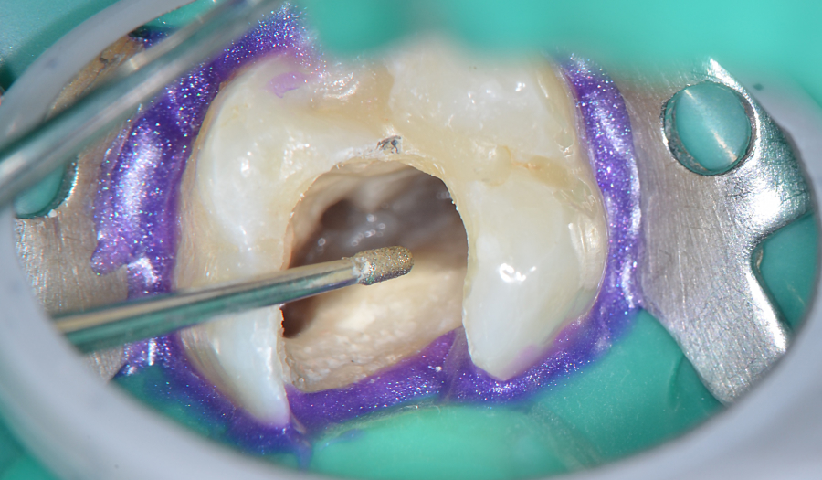
E6D ultrasonic insert used to locate the MB2.
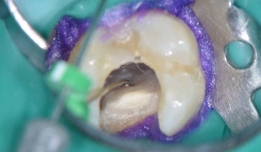
Manual file penetrating the MB2.
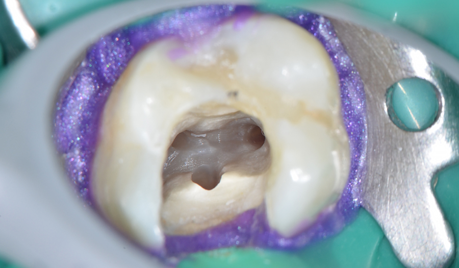
MB2 after location and preparation.

